

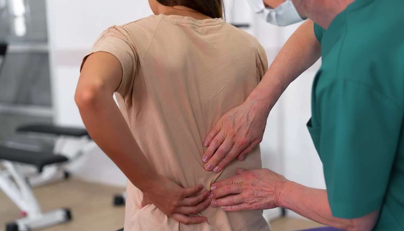
Your MRI Shows a Disc Herniation. But Is That Actually What’s Causing Your Pain?
You went to the doctor with back pain. Maybe the pain was shooting down your leg. The doctor ordered an MRI. The report came back with words like “disc herniation,” “disc protrusion,” “degenerative disc disease,” or “disc bulge at L4-L5.” And now you’re worried. The language sounds structural. It sounds permanent. It sounds like something that needs to be fixed.
Here is what most people are never told: disc herniations on MRI are incredibly common in people who have zero pain. Research from the Mayo Clinic reviewed over 3,100 MRI scans of people with no back pain at all. They found disc degeneration in 37% of 20 year olds and 96% of 80 year olds. Disc bulges were present in 30% of pain-free 20 year olds and 84% of pain-free 80 year olds. Disc protrusions showed up in 29% of 20 year olds who felt perfectly fine (Brinjikji et al., AJNR, 2015).
That means if you grabbed 10 random people off the street in Oakville who have never had a day of back pain in their lives, at least 3 of them would have disc bulges on MRI. They just don’t know it because they have no symptoms.
This does not mean your pain is not real. It absolutely is. But it means the disc herniation on the MRI report might not be the thing causing it. And understanding that distinction changes everything about how you approach treatment. At Wellmax Physiotherapy in Oakville, disc herniation MRI findings are something our physiotherapists assess and explain every week. The clinical examination, not the scan, determines the treatment plan.
Why MRI Findings Don’t Always Match the Pain
Disc Changes Are Part of Normal Aging
The spine ages just like the rest of the body. Disc degeneration is not a disease. It is a normal process that begins in your 20s and progresses throughout life. The same way your hair turns grey or your skin develops wrinkles, your discs lose hydration, develop bulges, and show wear on imaging. Calling this “degenerative disc disease” is misleading because it implies a pathological process. In most cases, it is simply your spine doing what spines do over time.
A landmark study published in the New England Journal of Medicine scanned the lumbar spines of 98 people who had never experienced back pain. Sixty-four percent of them had at least one abnormal disc finding. Twenty-eight percent had a disc bulge. The researchers concluded that these findings on MRI in people without symptoms must be interpreted cautiously because they are frequently incidental (Jensen et al., NEJM, 1994).
The Size of the Herniation Doesn’t Predict the Pain
Some people have massive disc herniations on MRI and moderate symptoms. Others have small bulges and severe pain. The relationship between the size of a disc abnormality and the amount of pain a person experiences is inconsistent. Pain is influenced by inflammation around the nerve root, the sensitivity of the nervous system, muscle guarding and spasm, movement patterns, stress, sleep quality, and a dozen other factors. A disc herniation may contribute to the picture, but it is rarely the entire picture.
This is why two patients with identical MRI reports can have completely different levels of pain and function. The scan shows structure. It does not show pain.
MRI Findings Often Don’t Change Even When Pain Goes Away
Here is something that surprises most patients. Many people recover completely from back pain or sciatica while their MRI still shows the same disc herniation. The herniation is still there on the scan. But the pain is gone. This happens because the initial pain was driven by inflammation, nerve sensitivity, and muscle guarding rather than by the disc pressing on a nerve. Once the inflammation settles and the nervous system calms down, the symptoms resolve even though the structural finding remains.
If the disc herniation were truly the sole cause of pain, the pain would only resolve when the disc went back to normal. But that is often not what happens clinically.
The Good News: Most Disc Herniations Shrink on Their Own
This is the part most patients never hear from their doctor. Multiple meta-analyses have now confirmed that the majority of disc herniations reabsorb without surgery. A 2017 meta-analysis published in Pain Physician pooled data from 11 studies and found an overall spontaneous resorption rate of 66.66% (Zhong et al., 2017). A more recent and larger meta-analysis in 2023 that included 31 studies and over 2,200 patients found an overall resorption rate of 70.39% (Zou et al., Clinical Spine Surgery, 2023).
Even more interesting: larger herniations actually tend to reabsorb faster than smaller ones. Disc sequestrations (where a fragment breaks off completely) had an 87.77% resorption rate. Disc extrusions had a 66.91% rate. The body treats the herniated material as foreign tissue and sends immune cells to break it down. This process takes weeks to months, and it happens naturally.
The resorption process mainly occurs within the first 6 months. This is exactly the window where physiotherapy is most effective at managing pain, maintaining activity, and preventing deconditioning while the body does its own healing work. Learn more about herniated disc treatment at Wellmax.
So When Does the MRI Actually Matter?
MRI is not useless. It serves an important purpose in specific clinical situations. The problem is when it is used as the first step rather than a targeted diagnostic tool. Here is when imaging genuinely changes the treatment plan:
Progressive Neurological Deficit
If weakness in the leg or foot is getting worse over days or weeks, that signals increasing nerve compression that may require surgical intervention. MRI confirms the location and severity.
Cauda Equina Syndrome
Sudden loss of bladder or bowel control, numbness in the groin or saddle area, or rapidly progressing weakness in both legs is a medical emergency. MRI is urgent in this scenario. Our back pain and sciatica page covers cauda equina red flags in detail.
Symptoms That Have Not Responded to 6 to 8 Weeks of Proper Conservative Treatment
If someone has followed a structured physiotherapy program with the correct exercises for their presentation, and they are not improving at all after 6 to 8 weeks, imaging helps determine whether a surgical consultation is the right next step.
Clinical Picture That Doesn’t Add Up
If the physical examination findings are inconsistent or suggest something beyond a typical disc or nerve issue, imaging helps rule out less common causes like infection, tumour, or fracture.
Outside of these situations, early MRI for back pain often does more harm than good. Research has consistently shown that patients who receive early MRI for uncomplicated back pain have worse outcomes, more surgery, higher healthcare costs, and longer disability periods than those who start with physiotherapy first. The scan itself can increase fear and avoidance behaviour, leading patients to stop moving because they believe their spine is “damaged.”
What Actually Works for Disc Herniations
Directional Preference Exercises
For most disc-related pain, there is a specific direction of movement that reduces symptoms. For many patients with disc herniations, extension (arching the back gently) centralizes the pain, meaning it pulls the leg pain back toward the spine. This is a positive sign that the nerve is decompressing. Finding and repeating this movement pattern multiple times per day is the single most powerful tool in early disc herniation management. Our back pain and sciatica page covers directional preference in detail.
Manual Therapy
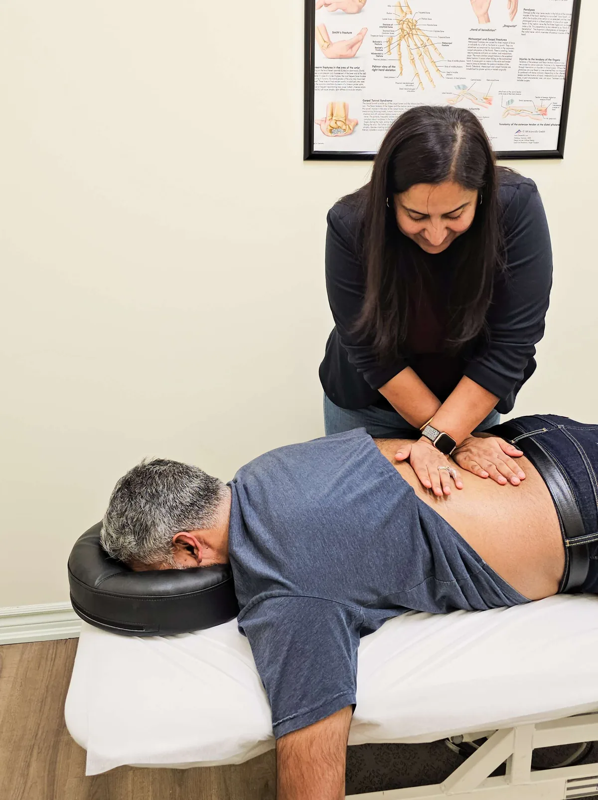
Hands-on joint mobilization and soft tissue work reduce pain, improve spinal mobility, and create a window for the patient to exercise more effectively. Manual therapy alone is not a long-term solution, but it accelerates the early stages of recovery and helps patients participate in their exercise program.
Progressive Core and Spinal Strengthening
Once the acute pain settles (typically within the first 2 to 4 weeks), the focus shifts to building spinal resilience. This means targeted strengthening of the deep core stabilizers (transversus abdominis, multifidus), the glutes, and the entire posterior chain. People who complete a structured strengthening program after a disc herniation episode have significantly lower recurrence rates than those who stop treatment once the pain fades.
Activity Modification, Not Bed Rest
Every major clinical guideline agrees: bed rest is harmful for disc herniations. Staying active, walking daily, and modifying activities (not eliminating them) is the evidence-based approach. Movement promotes disc nutrition, reduces inflammation, and prevents the deconditioning that makes recovery longer and harder.
Deep Tissue Laser for Acute Flare-Ups
Class IV deep tissue laser therapy reduces inflammation at the nerve root level during the first 2 to 3 weeks when pain is most severe. It is not a standalone treatment, but combined with directional preference exercises and manual therapy, it helps patients get through the worst of the acute phase faster.
When Surgery Is the Right Call
Physiotherapy resolves the majority of disc herniations. But honesty matters. Surgery is the right conversation when:
- There is progressive neurological deficit (worsening foot drop, spreading numbness, increasing weakness) that is not stabilizing with conservative care
- Cauda equina symptoms are present (this is an emergency, not a conversation)
- Severe pain has not responded to 8 to 12 weeks of proper, consistent physiotherapy
- The patient cannot participate in daily life or work despite appropriate conservative treatment
A microdiscectomy for the right candidate has high success rates and relatively quick recovery. The key word is “right candidate.” Surgery for patients whose pain is driven by central sensitization, fear avoidance, or muscle dysfunction rather than true nerve compression tends to produce disappointing results. The clinical assessment, not the MRI alone, determines who is a good surgical candidate.
At Wellmax, we are honest with patients about when conservative treatment has reached its limit. Being transparent about when to escalate is part of good care. We coordinate surgical referrals to orthopaedic and neurosurgical specialists when the clinical situation warrants it. Learn about our pre and post-surgical rehabilitation program.
What to Do If You Have Been Told You Have a Disc Herniation
Don’t panic. A disc herniation on MRI does not mean your spine is broken. It does not mean you need surgery. And it does not mean you will have back pain forever. The majority of disc herniations improve with time and the right management.
Get a proper clinical assessment. The MRI is one piece of the puzzle. A thorough physical examination tells us whether the disc finding is actually responsible for your symptoms, or whether something else is driving the pain. This distinction determines the entire treatment approach.
Start moving. Walking, gentle activity, and the right exercises prescribed by a physiotherapist will do more for your recovery than lying in bed waiting for the disc to “heal.” Your spine is designed to move. Movement is what keeps it healthy.
Commit to the strengthening phase. Pain relief is the first goal. But the strengthening program that follows is what prevents the problem from coming back. Skipping this phase is the most common reason disc herniations become a recurring issue.
Disc Herniation Treatment at Wellmax Physiotherapy in Oakville
Our physiotherapists see disc herniations every week. We understand the fear that comes with reading an MRI report full of clinical terminology. And we know that explaining what the findings actually mean, in plain language, is one of the most important parts of the first appointment.
We will assess your spine, test your neurological signs, find your directional preference, and give you a clear, honest answer about what is causing your symptoms and what needs to happen to resolve them. If the disc herniation is the issue, we treat it with directional preference exercises, manual therapy, progressive strengthening, and deep tissue laser when appropriate. If the disc is an incidental finding and something else is driving the pain, we treat that instead.
Expert care every visit. Direct billing to most major insurance providers. No referral needed.
Related Conditions and Services
Herniated Disc Physiotherapy, Comprehensive treatment for lumbar and cervical disc herniations at Wellmax.
Back Pain & Sciatica, Broader lumbar spine conditions including disc herniations, spinal stenosis, and chronic back pain.
Spinal Stenosis, Narrowing of the spinal canal causing nerve compression, leg pain, and walking limitations.
Chronic Pain, When back pain persists beyond normal healing timelines and involves central sensitization.
Deep Tissue Laser Therapy, Class IV laser for acute disc herniation flare-ups and nerve root inflammation.
Shockwave Therapy, Treatment for chronic conditions that haven’t responded to exercise alone.
Pre & Post-Surgical Rehab, Prehabilitation and recovery after microdiscectomy and spinal surgery.
Sports Physiotherapy, Return-to-sport programming after back injury resolution.
Book Your Assessment
If you have been told you have a disc herniation, if pain is shooting down your leg, if you are wondering whether you need surgery, or if you simply want a clear, honest explanation of what your MRI report actually means, get it assessed properly.
Wellmax Physiotherapy
1148 Winston Churchill Blvd, Unit B3, Oakville, ON L6J 0A3
Phone: (905) 829-1100
Email: info@wellmaxphysiotherapy.ca
Proudly Serving Our Local Communities:
- East Oakville: Winston Park, Clearview, and the QEW/403 Corridor.
- South Oakville: Eastlake, Maple Grove, and Old Oakville.
- North Oakville: Joshua Creek, Falgarwood, and Upper Middle Road.
- Mississauga Border: Clarkson, Sheridan Park, and Sheridan Homelands.
Insurance: Direct billing to most major providers. WSIB and MVA claims accepted. No referral needed.
Hours: Monday to Friday: 9am to 7pm | Saturday: 9am to 2pm | Sunday: 9am to 2pm (by appointment only)
Frequently Asked Questions About Disc Herniation and MRI
My MRI says disc bulge. Should I be worried?
It is worth taking seriously, but the wording on a report is usually more alarming than the finding itself. A Mayo Clinic review of over 3,100 scans of people with no back pain found disc bulges in 30 percent of pain free 20 year olds and 84 percent of pain free 80 year olds. The finding is common in people who feel fine. What matters is whether a clinical examination links that finding to your actual symptoms.
Does a bigger herniation mean more pain?
No, the relationship is inconsistent. Some people have large herniations on imaging and moderate symptoms. Others have small changes and severe pain. Pain is influenced by inflammation around the nerve root, how sensitive the nervous system is, muscle guarding, movement habits, stress and sleep. A disc finding can be part of the picture, and it is rarely the whole picture. Two people with matching reports can function completely differently.
Can a herniated disc heal on its own?
Most do improve. A 2017 meta analysis in Pain Physician pooled 11 studies and found a spontaneous resorption rate of about 67 percent. A larger 2023 review of 31 studies and over 2,200 patients found about 70 percent. Larger herniations tend to reabsorb faster than smaller ones. The body treats the herniated material as foreign tissue and breaks it down, mostly within the first six months, which is the window where physiotherapy helps most.
If my pain goes away, will the MRI change?
Often it does not, and that surprises people. Many people recover fully while the scan still shows the same finding. That happens when the pain was driven by inflammation, nerve sensitivity and muscle guarding rather than by the disc pressing on a nerve. Once those settle, symptoms can resolve with the structure unchanged. It is a good reason to judge your progress by what you can do, not by a repeat scan.
Do I need an MRI for my back pain?
Usually not as a first step. Imaging does not always tell the whole story, and research shows that people who get early MRI for uncomplicated back pain tend to have worse outcomes, more surgery and longer time off work than those who start with physiotherapy. Reading a frightening report can also make people stop moving. Imaging works best when it answers a specific clinical question, so start with an assessment.
When does an MRI actually matter?
In four situations. If leg or foot weakness is getting worse over days or weeks. If there is sudden loss of bladder or bowel control, numbness in the groin or saddle area, or fast weakness in both legs, which is a medical emergency and needs urgent care, not an appointment. If symptoms have not responded to six to eight weeks of proper conservative treatment. And if the examination does not add up and less common causes need ruling out.
Do I need surgery?
Most people do not. Surgery becomes the right conversation when nerve related weakness is getting worse and not settling with conservative care, when cauda equina symptoms are present, which is an emergency, when severe pain has not responded to eight to twelve weeks of proper physiotherapy, or when daily life and work are not possible despite good treatment. The clinical assessment, not the scan alone, decides who is a good candidate.
Should I rest in bed until it settles?
No. Every major clinical guideline agrees that bed rest is harmful for disc related pain. Staying active, walking daily and modifying activities rather than dropping them is the evidence based approach. Movement supports disc nutrition, reduces inflammation, and prevents the loss of conditioning that makes recovery slower. Your physiotherapist will tell you which activities to change and which to keep, so you keep moving without stirring things up.
How long before I start feeling better?
Acute pain typically settles over the first two to four weeks, and that is when the focus shifts to building strength. Six to eight weeks of proper conservative treatment is the usual checkpoint for whether the plan is working. Reabsorption of herniated material mostly happens within the first six months. Day to day ups and downs are normal, so judge progress by the trend over weeks rather than by one bad afternoon.
What does degenerative disc disease on my report mean?
The name is misleading. Disc changes begin in your 20s and continue through life, in the same way hair greys and skin wrinkles. In a 1994 New England Journal of Medicine study, 64 percent of 98 people who had never had back pain had at least one abnormal disc finding. The label describes a normally aging spine far more often than it explains why someone hurts. The examination is what sorts that out.
You Might Also Like


Gurmeet Rai, Registered Physiotherapist
Gurmeet is a Registered Physiotherapist with the College of Physiotherapists of Ontario and the Clinic Director and co-owner of Wellmax Physiotherapy in Oakville, with 16+ years of clinical experience. She focuses on complex, stubborn cases where earlier treatment has not worked, with advanced training in pelvic health, TMJ, vestibular, and sports and MSK rehab. Expect expert care from assessment to discharge, with honest guidance along the way. Read Gurmeet’s full profile or meet the team.
This article is for educational purposes only and does not replace a professional clinical assessment. Every body and condition is different. If you are experiencing pain or symptoms, book an assessment with a qualified clinician for advice specific to your situation. If you are experiencing a medical emergency, call 911 or visit your nearest emergency department.
